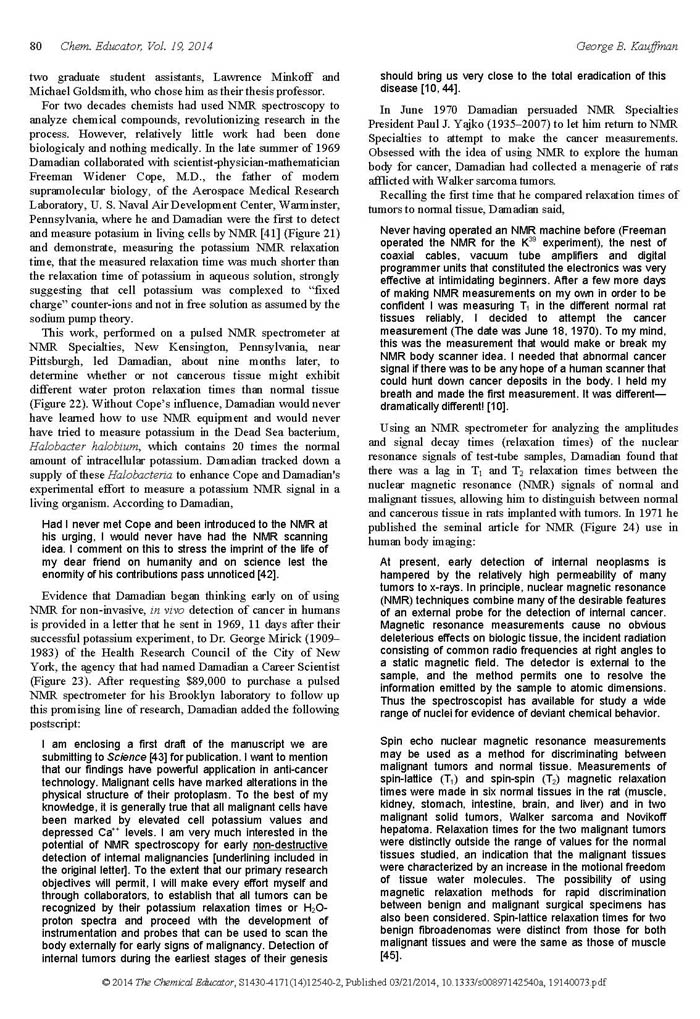
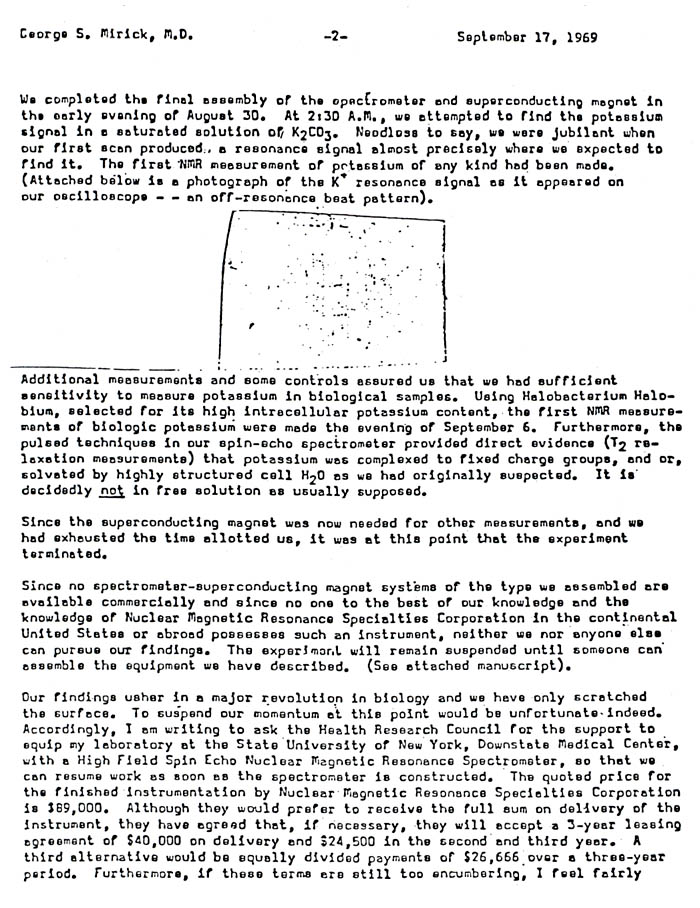
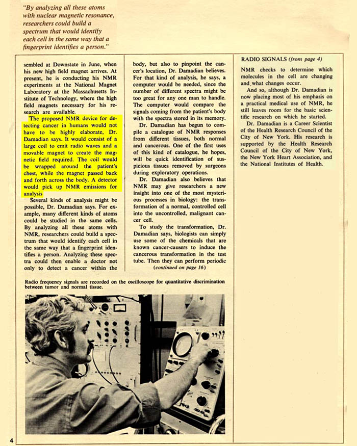
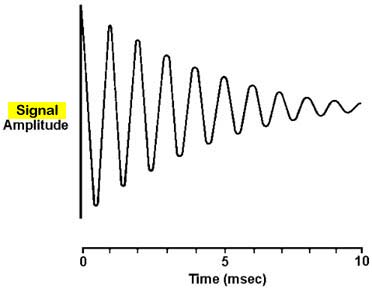
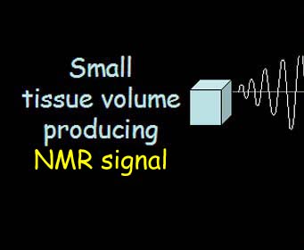
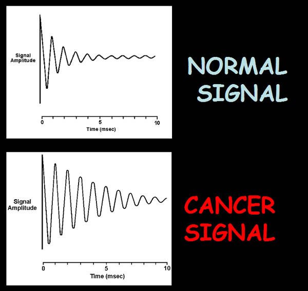
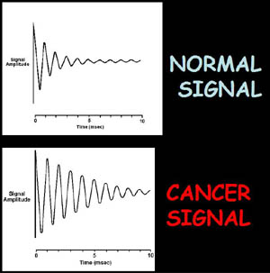
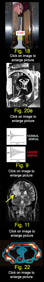
A live NMR signal such as that generated by a small tissue volume connected to an oscilloscope and an audio amplifier so that an example of an NMR signal that generates the MRI image can be directly visualized and heard. [ Click on Sine Wave to Listen ] THE STRENGTH OF THE SIGNAL1
SETS
THE PIXEL BRIGHTNESS ! 1. The computed strength of the signal (the amplitude) is determined by the signal's decay time (relaxation time). The longer the relaxation time the greater the signal amplitude and the greater the brightness of the picture elements (pixels) that compose the image. The Signal Makes The Image:
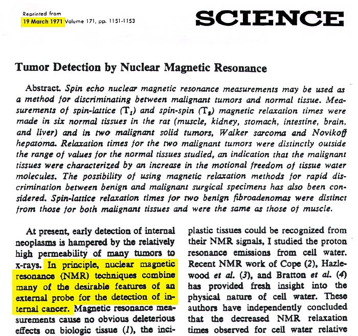
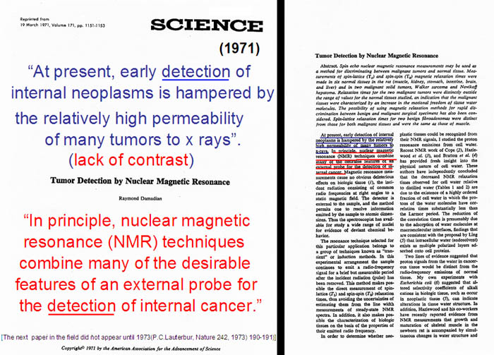
No Signal Differences1, No Image. 1. R. Damadian, Tumor Detection by Nuclear Magnetic Resonance. Science, 19 March 1971, Vol.171, pp. 1151-1153. Raymond V. Damadian is the medical doctor who first proposed scanning medical patients by NMR (nuclear magnetic resonance, the original name of the MRI) based on his DISCOVERY of the principle on which all modern MRI is based — the different NMR signals that tissues emit in a magnetic field. The amplitude of the signal determines the brightness of the picture element (pixel) that the MRI image is composed of. NO SIGNAL DIFFERENCES
AND ...
THE IMAGE IS A BLANK !!
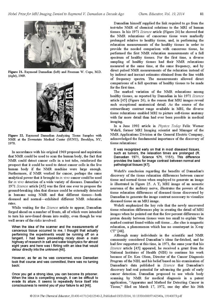
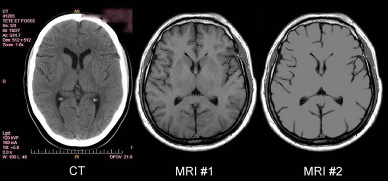
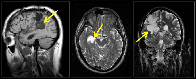
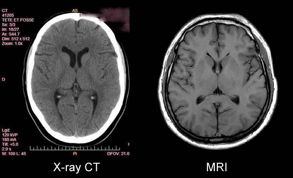
Note the soft tissue detail visualized in the MRI image #1 of the brain that is not visualized by x-ray CT technology (e.g. the pronounced white matter-grey matter differentiation of the MRI, the clearly defined thalamic nuclei, and the well visualized subdural layers not visualized by CT). MRI image #2 shows an image of the brain where all the MR signals of the brain tissue are the same. (i.e. no signal difference from the TISSUES of the BRAIN. No grey matter -white matter differentation, no caudate nucleus, no putamen, no thalamus.) In the absence of the MR signal differences of the normal tissues discovered by Damadian (Fig.6, Fig.9) the image detail of normal human anatomy is missing (MRI #2). NO SIGNAL DIFFERENCES:
THE IMAGE IS A BLANK THE SIGNAL MAKES THE IMAGE !!
NO SIGNAL DIFFERENCES: 1,
NO IMAGE !! AND
NO
ANATOMIC DETAIL
VISIBLE !!! (Fig 5 - MRI #2) Paul C. Lauterbur's Notebook
|
||||||||||||||||||||||||||||||||||||||||||||||||||||||||||||||||||||||||||||||||||||||||||||||||||||||||||||||||||||||


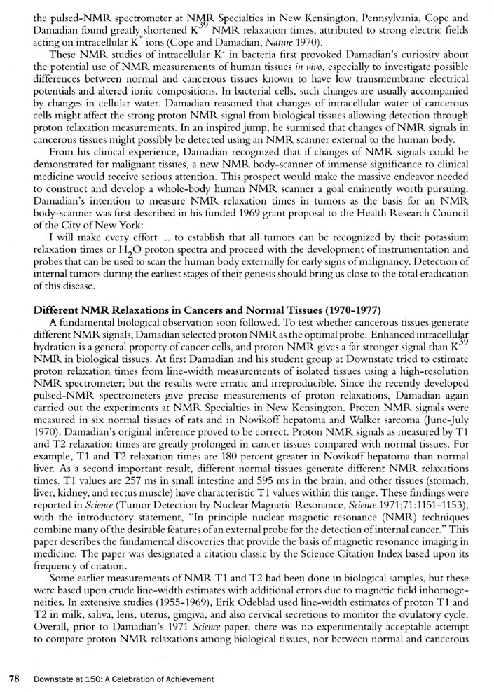
 Raymond
V. Damadian, MD, conceived the
Raymond
V. Damadian, MD, conceived the  “The
initial concept for the medical application of NMR,
as it was then called,
“The
initial concept for the medical application of NMR,
as it was then called, 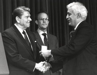 1.
1.
 3.
3.
|
T1 Image |
T2
Image |
T2 Image |
  |
||
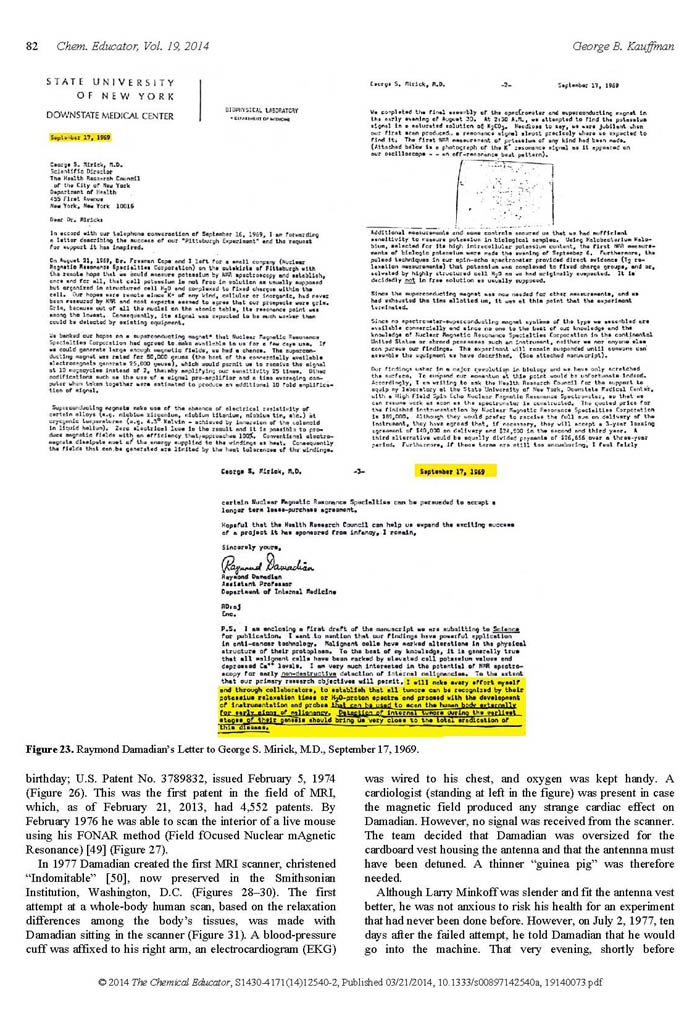
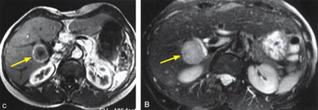
These signal amplitude differences enabled cancer tissues (Figures 11-13) and other tissues to be visualized in MRI images because the signal differences generate the needed brightness differences "PIXEL CONTRAST (IMAGE DETAIL)" in the picture elements (pixels) needed to visualize detail in the MRI image.
The CONTRAST
in pixel brightness, "PIXEL
CONTRAST (IMAGE DETAIL)", allows the cancer
pixels in the image to be distinguished from the surrounding
normal pixels. (Figs 11-13)
ALL THE IMAGE PIXELS
ARE EQUALLY BRIGHT !
INVISIBLE !!
The cerebellar tumor as it would appear (14-D) with no MR signal differences. Figure 14-D is the same image as Figure 14-B but where all MR signal differences were eliminated and all the MR pixels therefore had the same pixel brightness. The absence of the MR signal differences between cancer and normal tissue DISCOVERED BY DAMADIAN gives the MR image pixels equal brightness and
THE IMAGE IS A BLANK
NO IMAGE !!
INVISIBLE !!
(Fig 14D)
scanning for cancer?
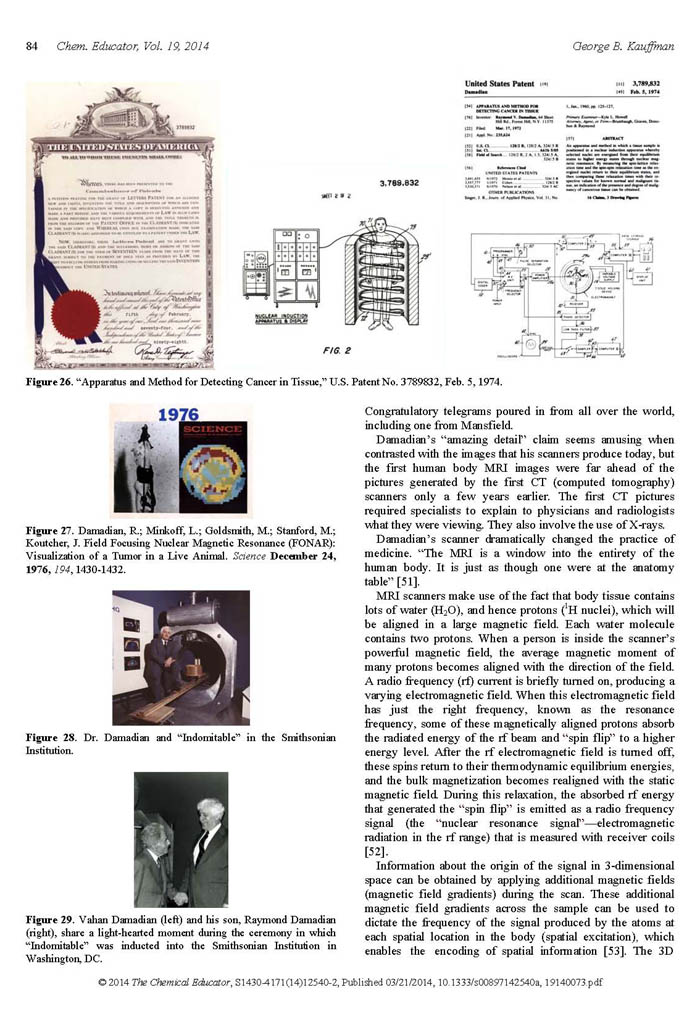
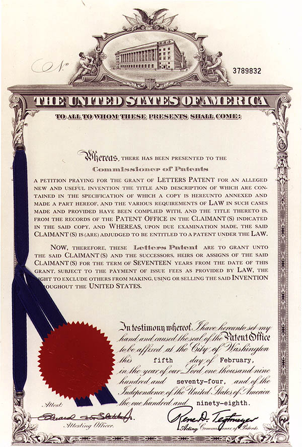
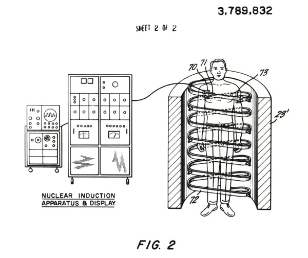
“Apparatus and Method
for Detecting Cancer in Tissue”
(the first ever patent on MRI)
“Apparatus and Method
for Detecting Cancer in Tissue”
Filed March 1972
 |
|
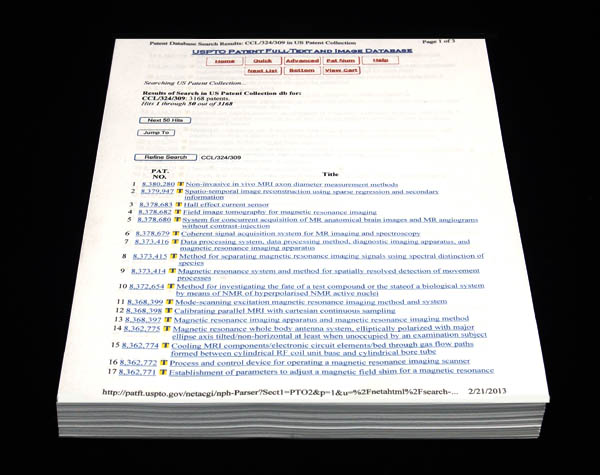
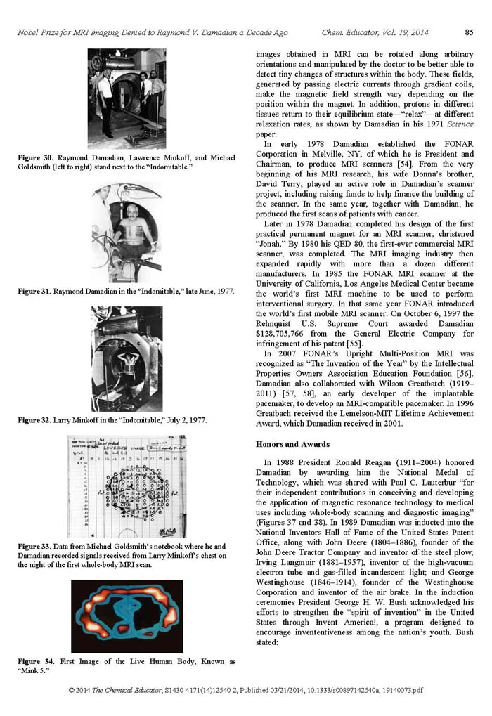
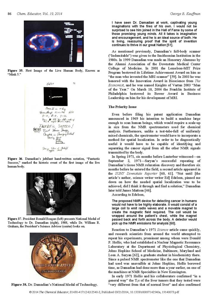
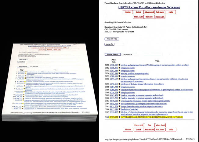
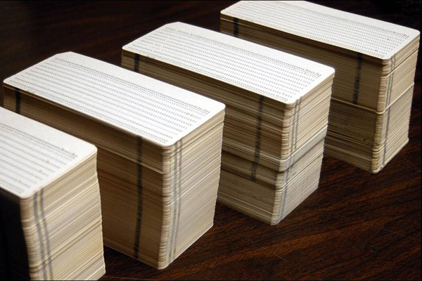
The above is the listing of all of the titles of the 4552 patents (as of 2/21/13) issued for MRI by the United States Patent Office following Dr. Damadian's original '832 patent for MRI (filed March 17, 1972).
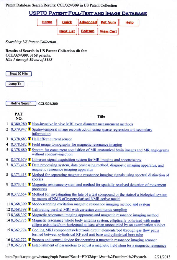
The above is the first page of the title list of the 4552 patents issued for MRI by the United States Patent Office beginning with the most recently issued patent (as of 2/21/13).
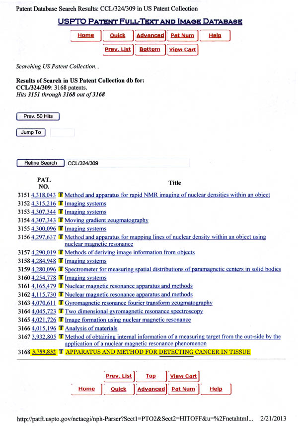
The conclusion of the title listing of the 4552 patents issued for MRI by the United States Patent Office (as of 2/21/13) following Dr. Damadian's original patent for MRI that inaugurated the MRI industry. [U.S. Patent "Apparatus and Method For Detecting Cancer in Tissue". #3,789,832. Filed March 17, 1972].
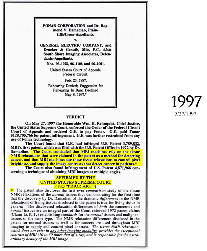
Upheld by the United
States Supreme Court
Oct 6, 1997
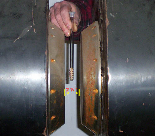
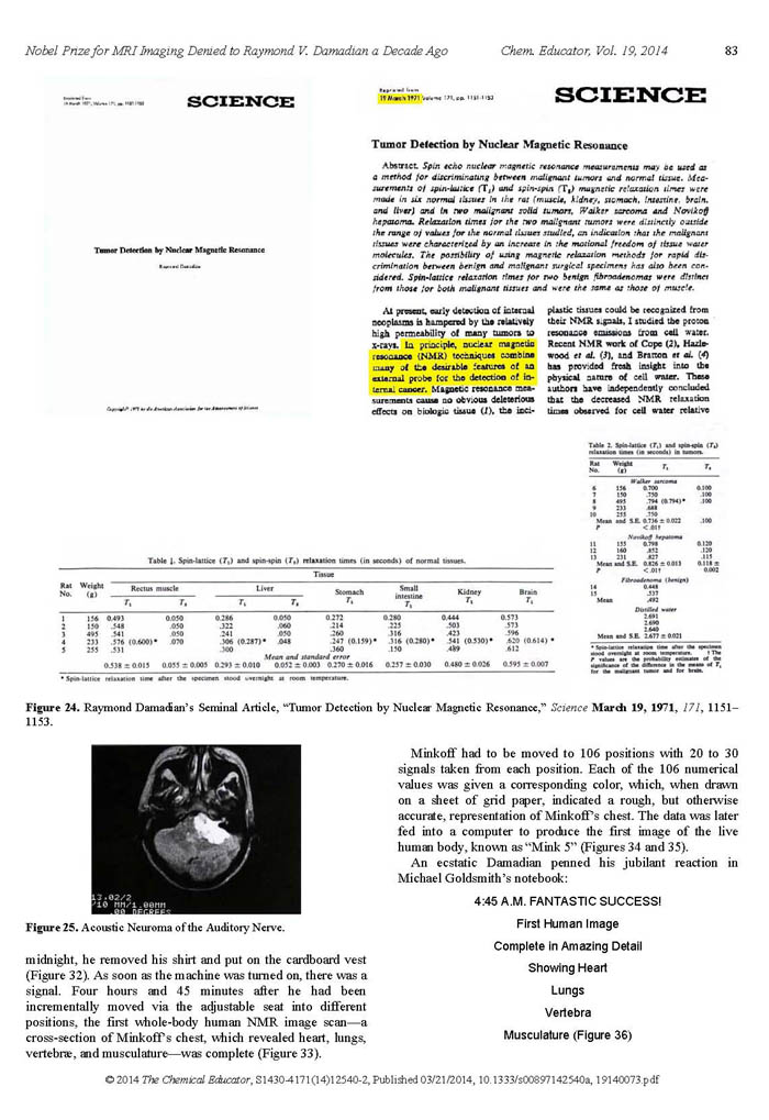
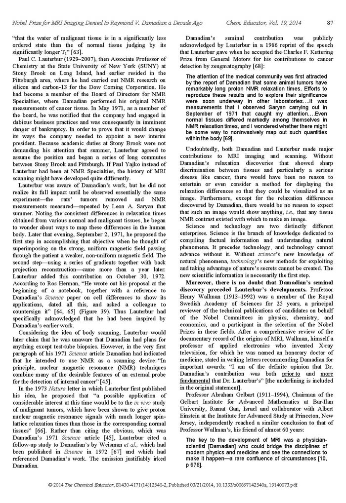
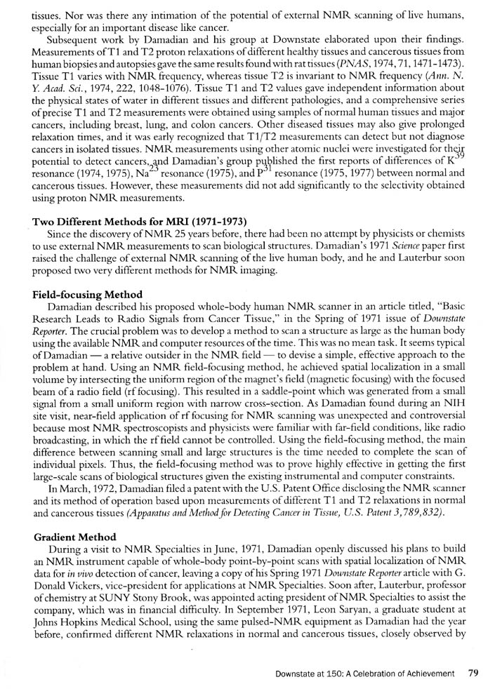
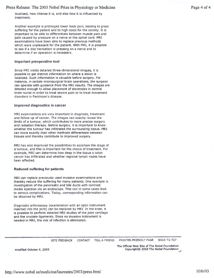
Figure
18.
The standard 23 year old 2 ¼"
NMR Test-Tube Analyser used by chemists
for ascertaining the molecule composition of aqueous
solutions prior to Dr. Damadian's discovery.
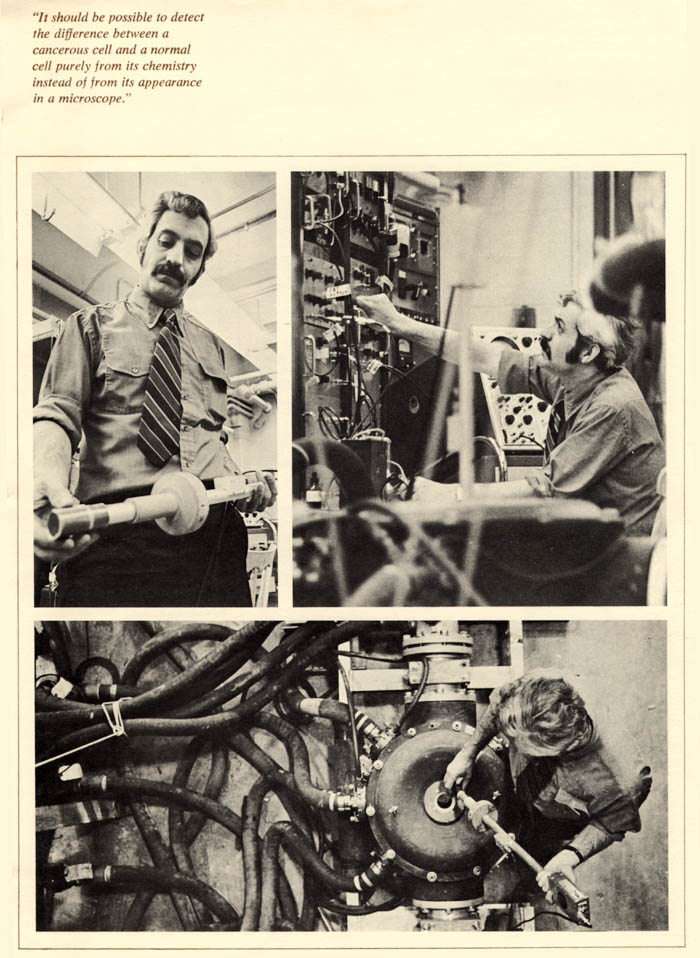
The standard NMR(MR) test-tube analyzer, utilized by NMR spectroscopists at the time had a two and 1/4 inch gap between the magnet poles to accept test-tube samples. It was the only NMR apparatus in existence at the time Dr. Damadian did his original NMR (MR) test-tube analyses of normal and cancerous tissue samples to see if a disease (cancer) differentiating NMR signal could be experimentally demonstrated that would enable his concept of a cancer detecting NMR(MR) body scanner to proceed.
Telling someone looking at this apparatus that it should be used to scan the human body was regarded as absurd. The giant magnets to do it did not exist. The rf antennas needed to accomplish detecting a less than 1mm tumor inside the body also did not exist. They were a major concern.
The sample tube is non-invasively wrapped with an external transmitter-receiver coil to stimulate and receive nuclear resonance signals from the sample.
With the same tissue sample as in the above illustration but now 10" removed from the proposed MR antenna envisioned for a body MR scanner, and where the MR signal itself was not all that strong and readily lost by the slightest mispositioning of the sample within the magnet the prospect of successfully acquiring an MR signal with an external antenna from a 1mm tissue sample deep within the human body was a major uncertainty.
At the time the idea (1971) of taking a 2¼ inch test-tube analyzer and turning it into a scanner of the live human body was deemed absurd.
“THEREFORE
ANY FURTHER DISCUSSION ABOUT SCANNING THE HUMAN BODY BY NMR IS VISIONARY NONSENSE ” |
| This
was the conclusion of an NMR scientist of the
John Hopkins Medical Center, one of the three
NMR scientists (Raymond Damadian, Carlton Hazlewood
and Donald Hollis) granted a research contract
by the National Cancer Institute's (NCI) Cancer
Diagnosis Project in 1976, after his successful
repeat of Dr. Damadian's demonstration of the
prolonged relaxations of the NMR signals of cancerous
tissues and his additional observations that non-malignant
diseased tissues
also had prolonged NMR relaxations. He had, however,
overlooked that both cancerous and non-cancerous
diseased tissue
NMR signals were markedly Professors of John Hopkins
Medical Center present at the conference immediately
disagreed with their colleague's "visionary
nonsense" claim stating that "Now Doctor,
just tell us where to put the needle we are way
ahead of where we are today". They further
refuted his declaration that it was "visionary
nonsense". |
Figure
19b. |
| At a subsequent and unrelated litigation the infringer of Dr. Damadian's patent made the same argument, that the elevated NMR relaxation times for cancer were also elevated in other diseased tissues that were non-cancerous. FONAR's attorneys responded, " Ladies and gentleman of the jury are you going to punish the guy because his original discovery detects MORE DISEASE than he originally envisioned ? " Dr. Damadian and Fonar prevailed. |
At a subsequent conference of NMR scientists where Dr. Damadian had been invited to present his NMR findings in cancer, the Moderator of the NMR conference, at the conclusion of Dr. Damadian's presentation stood to ask |
“ NOW DOCTOR HOW FAST DO YOU PROPOSE TO SPIN THE PATIENT ?”* |
* (Spinning the test tube sample at high rpm was a standard in NMR spectroscopy for overcoming the magnetic field inhomogeneities that the protons of the test tube sample were exposed to) |
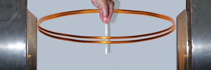
Using
Brookhaven National Laboratories magnet design software,
MAGMAP, that had been provided by the scientists from
Brookhaven National Laboratories, Dr. Gordon Danby,
Dr. Hank Hsieh and John Jackson, who had joined Dr.
Damadian as Fonar consultant employees to try to build
the first NMR (MRI) scanner of the live human body
that Dr. Damadian was trying to construct, Dr.
Damadian calculated that 30 miles (150,000 feet)
of SUPERCONDUCTING Niobium
Titanium (NbTi) magnet wire was needed to produce
the
5,000 gauss magnetic
field he had estimated was necessary to achieve a
successful NMR scan of a live human body. "At
the existing wire price of $1
per foot, the projected wire cost was $150,000
and I only had $15,000
in my budget"
Additionally the Niobium Titanium (NbTi) superconducting wire needed the making of SUPERCONDUCTING joints between successive lengths of wire in order not to undo the SUPERCONDUCTIVITY of the final magnet.
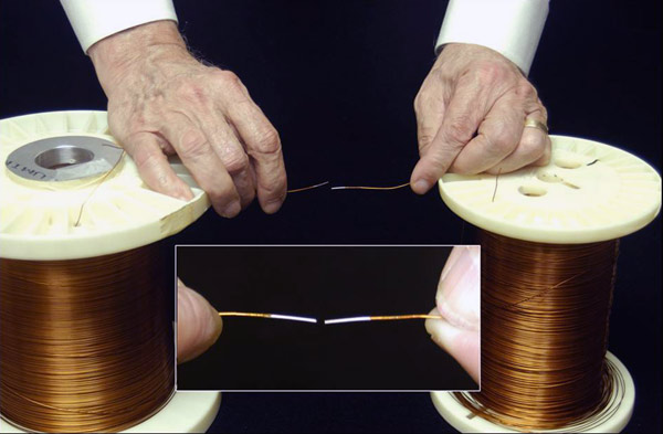
The NbTi liquid helium superconducting wire was not available from suppliers on a single spool. It required SUPERCONDUCTING joints to be made between successive lengths of the currently available NbTi wire.
I called Steve Lane, my sales representative at Westinghouse, the source of the SUPERCONDUCTING NbTi wire I was considering, and asked him if he would teach me how to make the superconducting joints I needed. His response was " What are you doing Dr. Damadian? Are you going into competition with Westinghouse? " I said " no no Steve I'm trying to make a superconducting NMR magnet that would be big enough to achieve an NMR scan of the live human body "
Steve responded by saying "it's good that you levelled with me Dr. Damadian. I can share with you something that no one else knows as yet. Westinghouse is going out of the business of making superconducting wire. I have about 30 miles of superconducting wire I can let you have for 10¢ on the dollar"
I was dumbfounded. With no prior knowledge from me, at Westinghouse or anywhere else, that I wanted to build an NMR magnet big enough to scan the human body, Westinghouse after 23 years of manufacturing their Niobium Titanium wire was SUDDENLY discontinuing their 23 year old manufacturing of NbTi wire, and had in their warehouse EXACTLY the amount of wire I had CALCULATED I NEEDED (30 miles) and would let me have it for THE EXACT AMOUNT I HAD IN MY BUDGET.
I was amazed by this extraordinary coincidence of Westinghouse ceasing to make the wire they had been making for 23 years at the precise instant I needed it, and at the exact length I needed (30 miles of wire) and at a price of " 10¢ on the dollar " that matched the exact amount I had in my budget ($15,000). I happened to mention this exceptional coincidence of the wire's sudden availability from Westinghouse at 10¢ on the dollar at the exact instant I needed it, to my wife's mother and father, Amy and "Bo" Terry (both evangelical christians).
My wife's mother responded " That's no coincidence Raymond " Ever since your Dad and I learned of your desire to build your scanning machine we've been praying for you " " This is not a coincidence. It's an answer to prayer ! "
From which I concluded that this sudden availability of the magnet wire AT 10¢ ON THE DOLLAR AT THE VIRTUAL INSTANT I NEEDED IT, was not an accident but that JESUS from my mother's prayers HAD JUST GIVEN MANKIND THE MRI !
" I WISDOM DWELL
WITH PRUDENCE AND FIND OUT KNOWLEDGE OF
WITTY INVENTIONS "
(Proverbs. 8:12 - KJV)
" FOR THE LORD GIVETH
WISDOM:
OUT OF HIS MOUTH COMETH KNOWLEDGE AND UNDERSTANDING
"
(Proverbs. 2:6–8 - KJV)
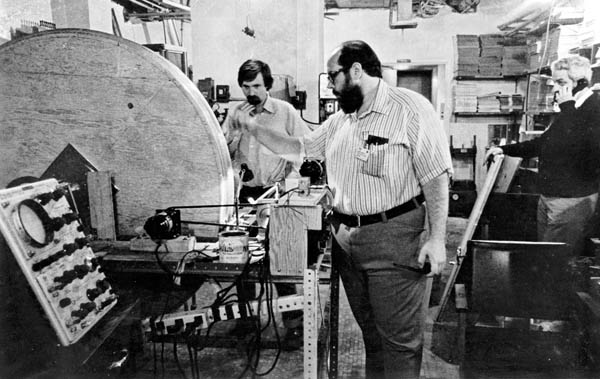
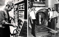
Figure 20a.
Michael Goldsmith and Michael Stanford winding one of the two Niobium Titanium (NbTi) superconducting magnet coils built for Indomitable.
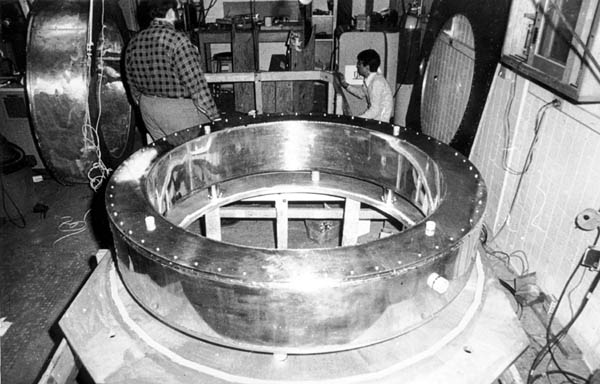
Figure 20b.
Michael Goldsmith and Nean Hu with the liquid helium cryogen chamber that housed the NbTi superconductiong magnet coil.
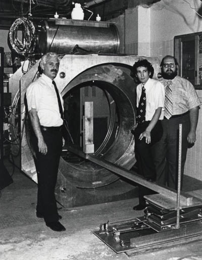
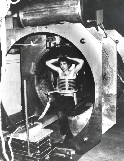
Left to Right, Raymond Damadian, Larry Minkoff and Michael Goldsmith alongside "live magnet" Indomitable with the iced liquid helium port on the top of the magnet alongside of the two helium cooling liquid nitrogen ports.
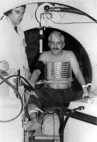
Figure 20d.
Dr. Damadian in Indomitable for the first attempt at a human MR scan with his chest surrounded by the largest diameter antenna (14" diameter) that Dr. Goldsmith had been able to build at the time that could still successfully generate any MR signal from an interior sample. Also pictured is an adjacent cardiac defibrillator to counter any emergencies that might arise and a cardiologist to administer it if necessary. The scan attempt on Dr. Damadian failed. All that was obtained was a normal EKG.
had just turned into a
SAD REALITY !
The Goldsmith hypothesis for the failed scan was Dr. Damadian was "too fat" for his coil and was loading the coil's impedance.
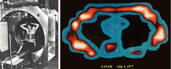
Larry Minkoff finally gets into Indomitable (weeks later) to test the Goldsmith "too fat" hypothesis.
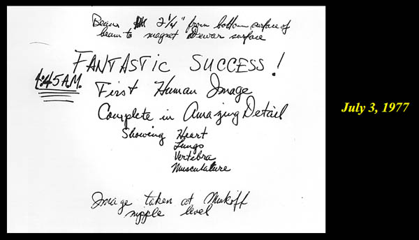
July 3, 1977

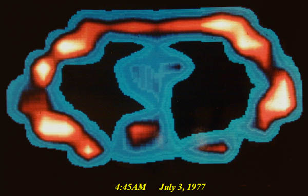
FIRST EVER MRI IMAGE OF THE LIVE HUMAN BODY !! A cross-section of L. Minkoff's chest at the level of T-8 showing chest walls, lungs, heart, aorta and vertebra, and the suggestion of cardiac chambers within the heart that was initially put down as too good to be true.
"VISIONARY
NONSENSE"
HAD JUST BEEN TURNED
into
REALITY !
The exhilaration of team Indomitable at 4:45 AM July 3, 1977.
WILD IDEA
( "Visionary Nonsense" )
( turning a 10 mm test tube analyzer, Fig. 18,
into a scanner of the LIVE human body )
HAD
MIRACULOUSLY
just become
of
23 years duration
had just been
Revolutionize Medical Imaging
DISCOVERED by Damadian
Revolutionize Medical Imaging
DETAIL
visualized in the
body's critical vital organs
for the first time in
medical history
(difference in pixel brightness of adjacent image pixels)
Fig. 14
(pixels-Fig 14c)
Accordingly
the power to visualize
DETAIL
in the body's
CRITICAL SOFT TISSUE VITAL ORGANS
(e.g. Brain, Heart, Muscles, Kidney, Liver, Spleen, Pancreas, Intestines)
therefore RESTS ENTIRELY on the power of the imaging technology to generate the
PIXEL CONTRAST
needed to visualize
IMAGE DETAIL
in the body's vital tissues. The existing x-ray technology for visualizing
IMAGE DETAIL
in the body's
CRITICAL VITAL ORGANS
had been severely lacking in its power to generate the
PIXEL CONTRAST
needed to visualize
IMAGE DETAIL
in the body's
CRITICAL
VITAL SOFT-TISSUE ORGANS
Dr. Damadian's
discovery
of the NMR signal differences of the body's vital
tissues, the signal amplitude differences generated
by their tissue NMR signal decay time differences
(their NMR "relaxation time" differences) provided
a
131% PIXEL CONTRAST
(Fig 6, Tables 1 & 2) for visualizing
IMAGE DETAIL
in the body's VITAL SOFT-TISSUE
ORGANS as compared to a maximum
4% PIXEL CONTRAST
available from X-ray.
This provided the
PIXEL CONTRAST
needed to visualize
IMAGE DETAIL
in the body's
CRITICAL
SOFT-TISSUE VITAL ORGANS
that had been missing from traditional medical imaging
(x-ray imaging)
for the better part of a century
(Roentgen 1895)
(Tables 1 & 2)
for the
FIRST TIME
IN MEDICAL HISTORY,
of the
CRITICAL SOFT TISSUE DETAIL
of the
LIFE GIVING
VITAL ORGANS
OF THE HUMAN BODY !!!
(Brain, Heart, Spine, Muscles, Liver, Kidney, Spleen, Pancreas, Intestines ...)
Damadian's discovery of the "signal that makes the image" provided another
VITAL DISCOVERY.
It provided,
FOR THE FIRST TIME IN MEDICAL HISTORY,
the power to achieve clear visualization of the body's
VITAL ORGANS.
Prior to the
advent of MRI, medical imaging from its x-ray inception
in 1895 was uniquely deficient in its ability to achieve
satisfactory (and necessary)
visualization of the life-sustaining organs of the
human body (Brain, Heart, Muscles,
Kidney, Liver, Spleen, Pancreas, Intestines ...).
X-ray technology was significantly limited in its
ability to produce the
PIXEL CONTRAST
needed to visualize
IMAGE DETAIL
in the body's soft-tissue vital organs due to the
small x-ray transmission differences of
x-ray radiation across the body's soft tissues (unlike
the transmission difference between bone and soft
tissue that generate pronounced image contrast on
x-ray images and excellent visualization of bone on
x-ray images). The difference in x-ray transmission
across the body's soft tissues, and therefore the
ability to generate the necessary PIXEL
CONTRAST in x-ray images to visualize
IMAGE DETAIL
in the body's
VITAL SOFT TISSUE ORGANS,
was severely limited to a maximum PIXEL CONTRAST of 4% by x-ray imaging technology.
The picture element (pixel) is the smallest display element in a medical image. A typical 256 x 256 pixel medical image is composed of 65,536 pixels (256 x 256). The ability to visualize detail in an image is dependent on the capability of adjacent picture elements (pixels) in an image to display differences in brightness (PIXEL CONTRAST) that reflect the differences in anatomy of the adjacent anatomic structures.
Consequently the ability of the pixels of an image to exhibit differences in brightness (PIXEL CONTRAST) and visualize ANATOMIC DETAIL in the image is dependent on the power of the imaging technology to generate differences in the brightness in the picture elements (pixels) that make up the image.
Accordingly
the magnitude of the PIXEL CONTRAST
achievable by the pixels
of an MRI image reflects the power of the image to
visualize ANATOMIC
DETAIL. Consequently the marked differences
in the T1 and T2 tissue NMR relaxations
(a 131% PIXEL
CONTRAST: Fig 6, Tables 1 &2)
achieved by MRI, as compared to the maximum of a
4% PIXEL CONTRAST
achieved by X-ray, accomplished
DETAILED VISUALIZATION
of
the body's
CRITICAL
LIFE-GIVING VITAL ORGANS
(Brain, Heart, Muscles, Kidney, Liver, Spleen, Pancreas,
Intestines ...)
FOR THE FIRST TIME
IN MEDICAL HISTORY
THE EXCEPTIONAL ANATOMIC DETAIL
ENABLED
by
THE TISSUE NMR RELAXATION
DISCOVERIES
of
Dr.
Damadian
that was UNPRECEDENTED
IN MEDICAL HISTORY and
had been beyond reach in medical imaging for nearly
a century (Roentgen 1895).
UNPRECEDENTED
IMAGE DETAIL
WITH NO
IONIZING RADIATION !
Figure 8.
T1 and T2 Medical Images of the Brain
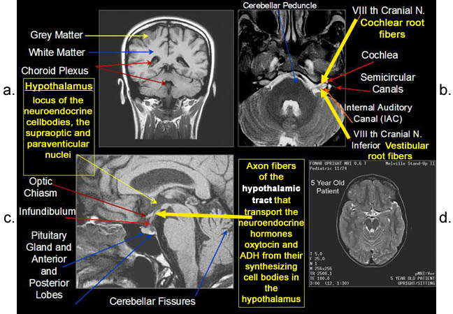
Figure 8a-8d. Further examples of
the exceptional anatomic detail made visible by
the DISCOVERY
of Damadian of the pronounced differences in the
decay rates (relaxations) of the NMR signals
of the body's normal tissues (Figure
6). The DISCOVERED
differences supply the pixel amplitude differences,
"PIXEL CONTRAST (IMAGE DETAIL)",
that produce, for the first
time in medical history, the detailed visualization
of normal human anatomy MRI is noted for. Note the
visualization of the
vestibular and cochlear nerves
WITHIN
the internal auditory canal (Figure 8b) and the
visualization of the
hypothalamic tract (that transports
hormones from the brain) WITHIN
the pituitary stalk. (Figure 8c)
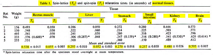
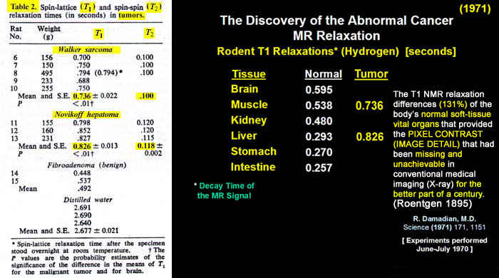
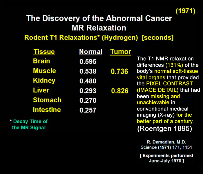
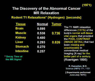
Damadian's
discovery
of the NMR signal differences that provided the MRI
visualization and detection of diseased tissue (Fig.
6, Tables 1 & 2) overcame this deficiency also. He
discovered
that the NMR signal
differences (T1
and T2 signal relaxation differences) among
the body's NORMAL soft
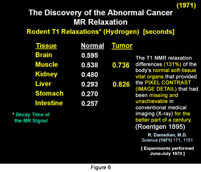
tissues (Brain, Muscle, Kidney, Liver, Stomach,
Intestine) were also pronounced
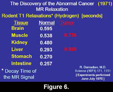
(Fig. 6, Tables 1 & 2) (e.g. small intestine 257msecs,
brain 595 msecs, a 131%
difference, Fig. 6). These large
NMR signal differences in the body's NORMAL
soft tissues, discovered
by Damadian, overcame, for the first time,
medical imaging's longstanding deficiency in its visualization
of the body's
VITAL
soft tissue normal organs (Figs. 7, 8).
The newly discovered
marked differences in
the proton NMR relaxation
times of the body's VITAL
soft tissues (131%)
enabled medicine to SURMOUNT
its historic deficiency, it's inability to generate
the necessary image
CONTRAST (PIXEL
CONTRAST)
crucial to visualizing the needed ANATOMIC
DETAIL of the body's
CRITICAL LIFE-GIVING VITAL ORGANS.
As Dr. Felix Wehrli PhD, MRI Imaging scientist and manager of the NMR Applications Division of the General Electric Company in his 1992 publication (Wehrli, F.W. Physics Today, June 1992, 34-42) REPORTED
" The Origins and Future of Nuclear Magnetic Resonance Imaging "
(regarding
the fundamental importance of the discovery
by Dr. Damadian
of the abnormal NMR relaxation times of diseased tissue),
" it was recognized early on
that in most diseased tissues, such as tumors,
the relaxation times are prolonged
(R. Damadian, 1971, Science 171, 1151)."
"This difference provides the basis for image CONTRAST between normal and pathological tissues ".
(Wehrli, F.W. Physics Today, June 1992, p38)
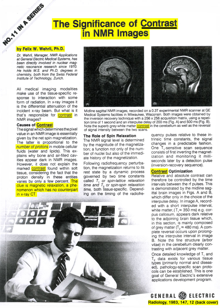
Wehrli had previously reported
in
" THE SIGNIFICANCE OF CONTRAST IN NMR IMAGES ",
that
"
the number of protons... does
not explain the marked CONTRAST
found within soft tissue, considering the fact that
the proton density in these entities varies by only
a few percent.
THE CLUE IS
magnetic
relaxation,
a phenomenon which has no counterpart in x-ray
CT "
(Wehrli, F.W. Radiology 1983, 147, 12 (back cover).
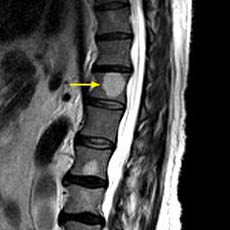
of all MRI images acquired today
T1 weighted or T2 weighted MRI images
(e.g. T1 axial lumbar MRI, T2 coronal brain MRI)
CHANGES
in the
T1 and T2 NMR SIGNALS OF THE BODY'S TISSUES that are created by the pathologic tissue changes generated by disease.
existed
NOWHERE
in the
PRACTICE OF MEDICINE
until
AFTER
Dr. Damadian's
DISCOVERY
of the
ALTERATION of T1 and T2
by
DISEASE
and their
POWER
for
DETECTING DISEASE
in the
BODY'S LIVE TISSUES ! :
PIXEL CONTRAST
(IMAGE DETAIL)
in images of the body's soft-tissue vital organs that had been missing and unachievable in conventional medical imaging
for the better part of a century
(x-ray).
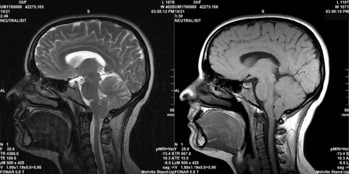
T2 sagittal MRI image of the brain T1 sagittal MRI image of the brain
DISCOVERY
of the
ABNORMALITIES
of the
T1 and T2
TISSUE NMR SIGNALS
GENERATED
by
TISSUE DISEASE !
|
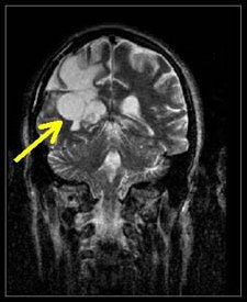
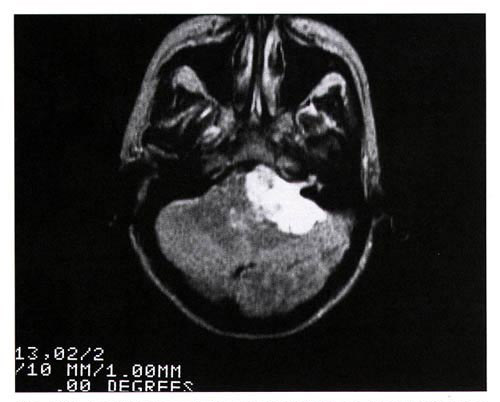
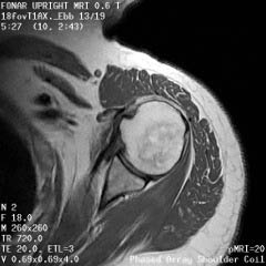
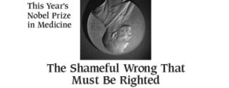
Figure 11.
T2 MRI visualization of a tumor of the brain made possible
by the discovery
of Damadian of the abnormal T2 (and T1) MR (NMR) relaxations
of cancerous tissue.
QUESTION:
What did MRI bring to medical
imaging that had been lacking
for the better part of a century
(W. Roentgen X-ray 1895) ?
ANSWER:The power to visualize
Unprecedented
Medical Image Detail in a medical image
for the first time in medical history.
(Fig 7 & 8)
 Figure
7
Figure
7
Figure 8
QUESTION: What did Dr.
Damadian's DISCOVERY
provide to medical images that SURMOUNTED
the inability of existing medical imaging technology
(x-ray) to visualize DETAIL
in medical images?
ANSWER: PIXEL CONTRAST
QUESTION: What delivered
the
PIXEL
CONTRAST ?
ANSWER: T1 and T2.
Picture Elements (PIXELS):
the structural component of the medical image
A typical MRI image is composed of 65,536 PICTURE
ELEMENTS (PIXELS), i.e. a rectangular 256 X
256 PIXEL MATRIX (Fig
10) consists of 256 pixel rows and 256 pixel columns.
![]()
Fig 10.
The power to visualize detail in any pixel image resides
in the power of the individual image pixel to generate
differences in pixel brightness (PIXEL
CONTRAST)
(Fig 14a, b, c, d)
![]()
(Fig 14a, b, c, d)
The differences in the tissue
NMR relaxation times (T1 and T2) of the body's
healthy tissues (131%),
DISCOVERED
by Dr. Damadian (Tables 1 & 2, Fig 6), provided
the signal amplitude differences that generate the
pronounced brightness DIFFERENCES
of the MRI image pixels and produce a 131%
PIXEL CONTRAST for the visualization of ANATOMIC
DETAIL in MRI medical images that had been
limited to a maximum PIXEL
CONTRAST of 4% for visualizing anatomic
detail by x-ray.
IMAGE DETAIL achieved by the MRI.
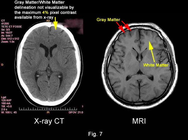
Note how the 131% PIXEL CONTRAST achieved by the MRI as compared to the maximum 4% PIXEL CONTRAST provided by x-ray visualizes and distinguishes the GRAY MATTER OF THE BRAIN FROM THE WHITE MATTER OF THE BRAIN (Fig 7) which cannot be achieved by the maximal 4% PIXEL CONTRAST available from x-ray.
DISCOVERED by Dr. Damadian
(Fig 6, Tables 1 & 2)
PRONOUNCED NMR signal differences
body's CRITICAL TISSUES
(Fig 6, Tables 1 & 2),
life-sustaining soft tissue vital organs (brain, heart, liver, spinal cord, intestines, etc)
visualized in detail in a medical image.
FIRST TIME IN MEDICAL HISTORY !!!
DISCOVERED by Dr. Damadian
(Fig 6, Tables 1 & 2), supply the missing anatomic detail that had been lacking in medical images
for the better part of a century
(Roentgen 1895)
 |
 |
 |
 |
 |
 |
 |
 |
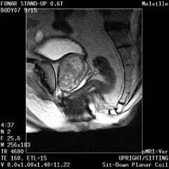
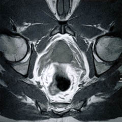
The Prostate without an Endorectal Probe |
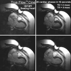
The Heart |
 |
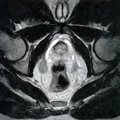 |
| Prostate: Delineation of Peripheral Zones (PZ), Cortical Zone (CZ) and Vescicles without an Endorectal Coil. | |
 |
 |
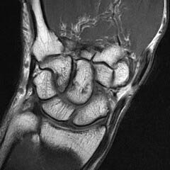
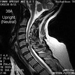
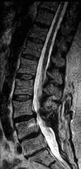
Cervical Spine |
|
Upright Neutral |
Upright Extension |
Unsuspected
Disc Herniation in Extension |
|
 |
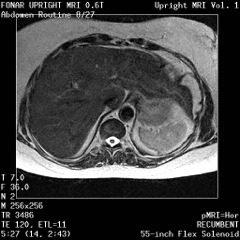 |
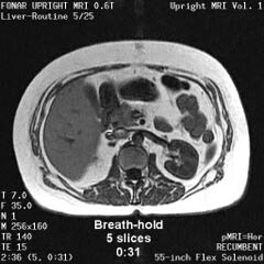
The Liver, Kidney
and Small Intestine
|
|
 |
|
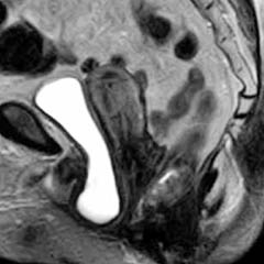
Bladder
and Uterus in Pelvic Floor Dysfunction (PFD) |
|
 |
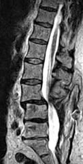 |
Lumbar Spine |
|
Recumbent, Weightless |
Upright, Weight-Bearing |
Figure 8.
Figure 8a-8d.
Further examples of the exceptional anatomic detail
made visible by the DISCOVERY
of Damadian of the pronounced differences in the decay
rates (relaxations) of the NMR signals
of the body's normal tissues (Figure
6). The DISCOVERED
differences supply the pixel amplitude differences
"PIXEL CONTRAST (IMAGE DETAIL)"
that produce, for the first time in medical history,
the detailed visualization of normal human anatomy
MRI is noted for. Note the visualization of the
vestibular and cochlear nerves
WITHIN
the internal auditory canel (Figure 8b) and the visualization
of the hypothalamic
tract (that transports hormones from
the brain) WITHIN
the pituitary stalk. (Figure 8c)
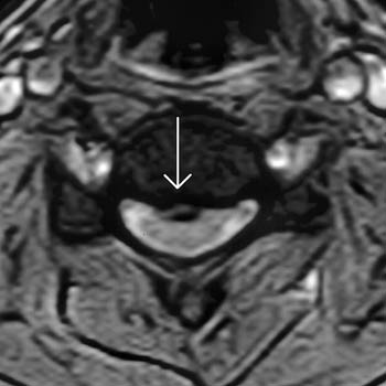
Lumbar Disc Herniation
Figure 24.
Merril Simon - Co-author
" The Pioneers of NMR and Magnetic Resonance
in Medicine - The Story of MRI", James Mattson
and Merrill Simon
Bar-Ilan University Press, 1969
of the Live Human Body 7/3/1977
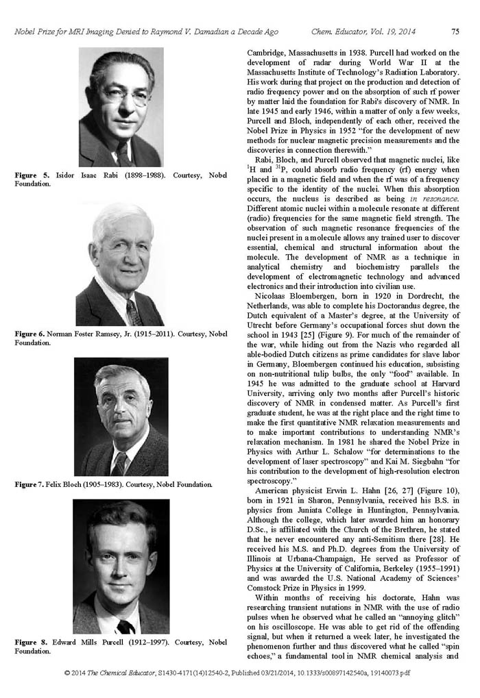
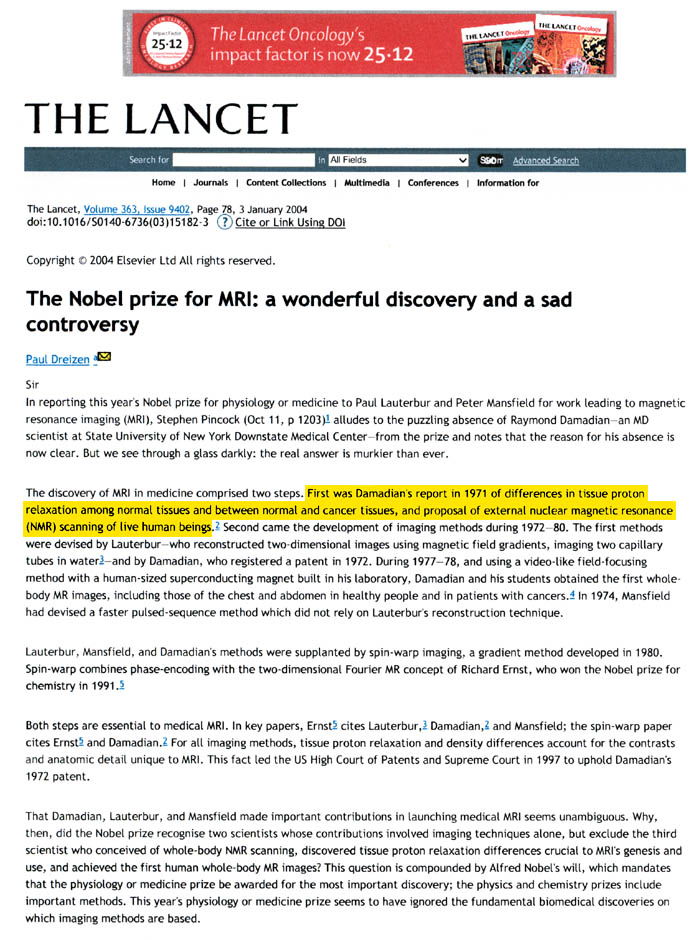
"We are perplexed, disappointed and angry about the uncomprehensible exclusion of Professor Raymond Damadian M.D. from this year's Nobel Prize in Physiology or Medicine. MRI's entire development rests on the shoulders of Damadian's discovery of NMR proton relaxation differences among normal and diseased tissues and his proposal of external scanning of NMR relaxation differences in the human body, published in Science in 1971"Eugene
Feigelson, M.D. |
Figures
30a-30c. First ever MRI images of patients with cancer
(1978)
(obtained on Indomitable)
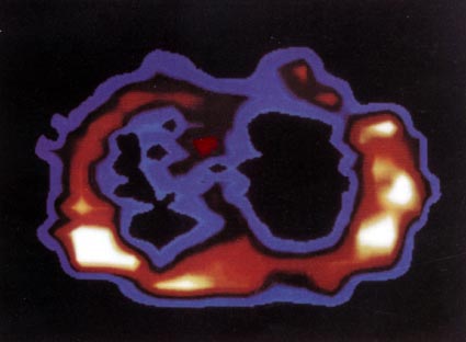 |
Figure 30a.
FONAR scan at the level of 1-3/4 inches below
the Angle of Lewis, by
the method of Indomitable1,
(Figs 20c and 20e)
in a man 46 years old with pulmonary oat cell
carcinoma. Tumour indicated by light blue infiltrate
in left lung field, which should be black as it
is in right lung cavity. Midline structure (red)
separating the two lung cavities is the cross
section through the arch of the aorta. |
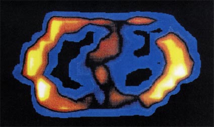 |
Figure 30b. FONAR cross-sectional
scan through the thorax at the level of the 3rd
intercostal space, by
the method of Indomitable1,
(Figs 20c and 20e)
in a patient with an adenocarcinoma of the breast
that metastasized to the right lung. The tumor
is seen as a band of signal-producing
tissue (light blue) bridging the right lung cavity.
The tortuous structure separating the right and
left lung cavities is the aortic arch. (1978)
(Scanning time: 36 min.) |
 |
Figure 30c. FONAR cross-sectional
scan through the low chest (10th thoracic vertebra),
by the method of Indomitable1,
(Figs 20c and 20e)
in a patient with advanced alveolar cell carcinoma.
The tumor is seen as intense signal-producing
tissue (red, and less signal-intense
light blue) invading both lung cavities and obliterating
the bulk of the air space. (1978) (Scanning time:
30 min.) |
First Commercial MRI Company Founded
FONAR Corporation
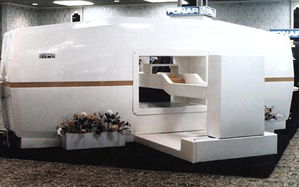 |
Figure 31. First-ever commercial MRI the FONAR QED 80. The FONAR QED 80 was equipped with a computer driven patient transport system (Vertical white bed mount pictured at the front of the QED 80 scanner) to automate the manual three-dimensional step-wise scanning procedure utilized by Indomitable1. (Indomitable transport apparatus Figs. 20c & 20e) |
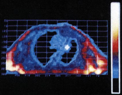 |
Figure 32. "Carcinoma
of the left upper lobe with peripheral consolidation".
Images published by Drs. Ross, Lie, Thompson &
Associates from their FONAR QED 80 MRI scanner
installed in their radiology practice in Clevelend,
Ohio. QED 80 images were acquired by the step-wise
3D patient translocation method of Indomitable1. |
1. The 3D step-wise translocation of the patient across the magnetically focused resonance aperture. The resonance aperture was achieved by focussing the "near field" magnetic component of the transmitted rf (U.S. Patent 3,789,832) in combination with the shaping of the static magnetic field of the region of interest to generate a spatially localized NMR signal. |
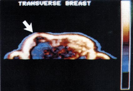 |
Figure 33. "NMR
image of the breast shows a large mass (dark area)
in the central portion of the right breast. T1
data are consistent with the diagnosis of cyst.
(mean: 151, width: 239)"
Images published by Drs. Ross, Lie, Thompson &
Associates from their FONAR QED 80 MRI scanner
installed in their radiology practice in Clevelend,
Ohio. QED 80 images were acquired by the step-wise
3D patient translocation method of Indomitable1. |
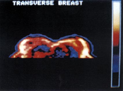 |
Figure 34. "NMR
image shows a region of low density in the left
breast with elevated T1 values. The small black
area in the right breast is compatible with the
diagnosis of cyst. (mean: 127, width: 128)"
Images published by Drs. Ross, Lie, Thompson &
Associates from their FONAR QED 80 MRI scanner
installed in their radiology practice in Clevelend,
Ohio. QED 80 images were acquired by the step-wise
3D patient translocation method of Indomitable1. |
“God hath made man Upright”
Ecclesiastes 7:29
He Was Made ?
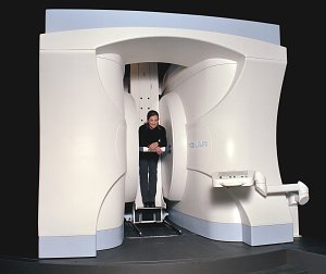
The Upright MRI Begins
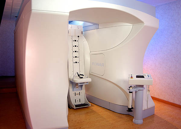
Figure 35.
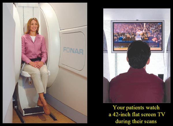
Figure 36.
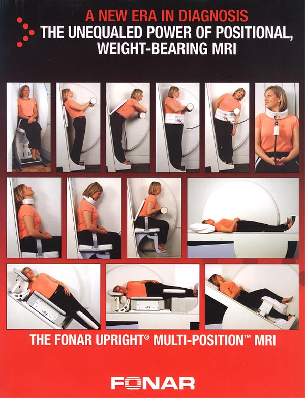
Figure 37.
Mommy and Me MRI
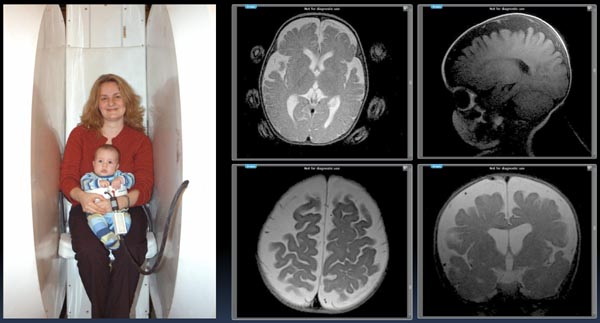
Figure 38.
The Mommy and Me MRI
The Mommy and Me MRI produced the above (just obtained) infant pictures of a 7 month old child WITHOUT ANESTHESIA with the infant lying in the scanner in a Fonar receiver coil with the mother kneeling and facing into the scanner (opposite to the position shown) holding the child's head. The upper left image of the infant's brain shows the mother's head positioning finger-tips. The brain images obtained exhibit hydrocephalus in the infant together with pronounced CSF pooling suggestive of significant obstruction to the flow of CSF (most likely cervical obstruction) in and out of the brain generating increases in intracranial pressure (ICP) and cerebral pooling of CSF as visualized in the above brain images of the infant.
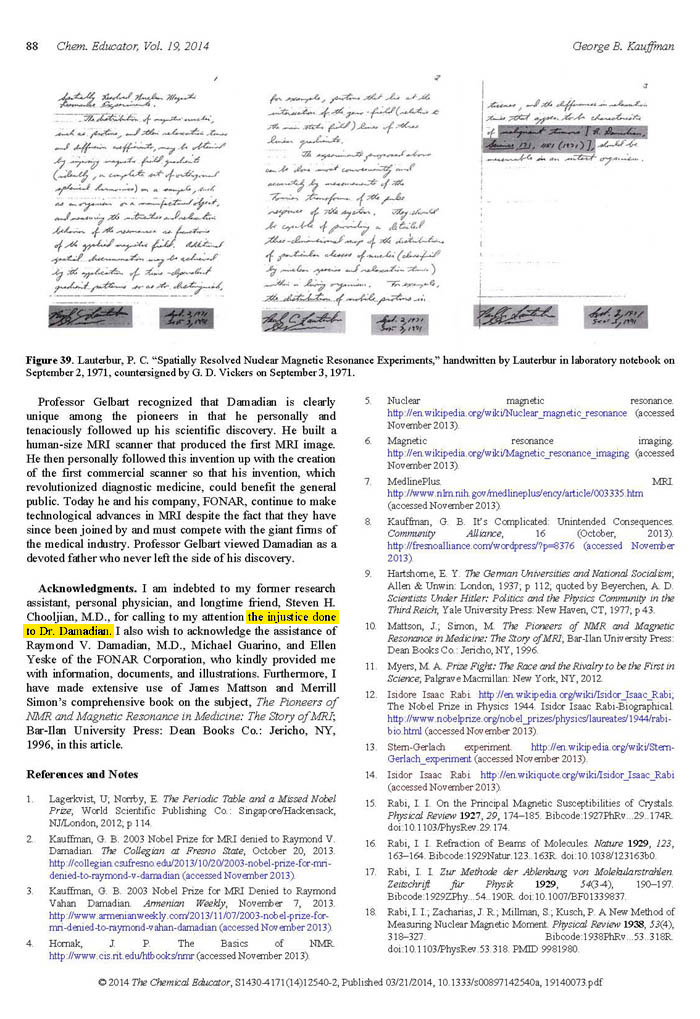
Figure 39.
THE TRUTH OF HISTORY, THE UNIVERSITY OF CHICAGO PRESS – 2 YEARS AFTER THE NOBEL PRIZE
 “By
the final few decades of the twentieth century, medical
practitioners were exploiting developments in nuclear
physics to provide a range of new ways of peering
inside the human body …. Another technique developed
during the 1970s was MRI (magnetic resonance imaging).
The technique was initially developed by Raymond Damadian
(1936 -), working at the Downstate Medical Center
in New York, making use of the fact that different
atomic nuclei emit radio waves of predictable frequencies
when exposed to a magnetic field. Damadian
noted that tumorous cells emitted signalsdifferent
from those emitted by healthy tissue and used this
as the basis for a new technique for identifying cancers.
Damadian and his fellow workers produced the first
MRI scan of the human body in 1977.”
“By
the final few decades of the twentieth century, medical
practitioners were exploiting developments in nuclear
physics to provide a range of new ways of peering
inside the human body …. Another technique developed
during the 1970s was MRI (magnetic resonance imaging).
The technique was initially developed by Raymond Damadian
(1936 -), working at the Downstate Medical Center
in New York, making use of the fact that different
atomic nuclei emit radio waves of predictable frequencies
when exposed to a magnetic field. Damadian
noted that tumorous cells emitted signalsdifferent
from those emitted by healthy tissue and used this
as the basis for a new technique for identifying cancers.
Damadian and his fellow workers produced the first
MRI scan of the human body in 1977.”
(Making Modern Science, A Historical
Review, The University of Chicago Press, 2005).
THE TRUTH OF HISTORY, SUNY HEALTH SCIENCES CENTER (DOWNSTATE MEDICAL CENTER), BROOKLYN – 5 YEARS AFTER THE NOBEL PRIZE
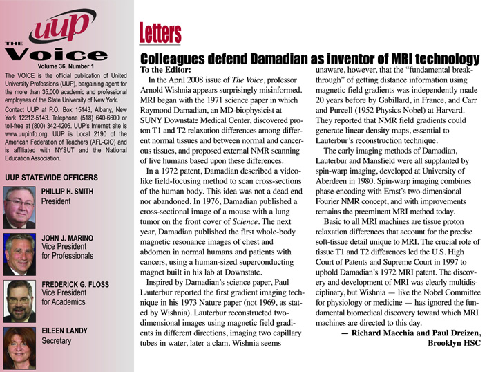
Figure 40.
(Richard Macchia, MD, and Paul Dreizen, MD, in the UUP Voice, the official publication of United University Professions (UUP), State University of New York.)
THE TRUTH OF HISTORY, THE AMERICAN INSTITUTE FOR MEDICAL AND BIOLOGICAL ENGINEERING – 6 YEARS AFTER THE NOBEL PRIZE
American Institute for Medical and Biological Engineering (AIMBE)Washington, D.C.
Figure 41.
Figure 42.
IN SUMMATION,
ORIGINATION OF THE NMR BODY SCANNING IDEA
Damadian both originated the IDEA to scan the human body by NMR (MR) and provided the means (the NMR signal differences) to achieve it. For any scientific advance to occur, someone must generate the IDEA to bring it about. Damadian provided the IDEA to give rise to the MRI.
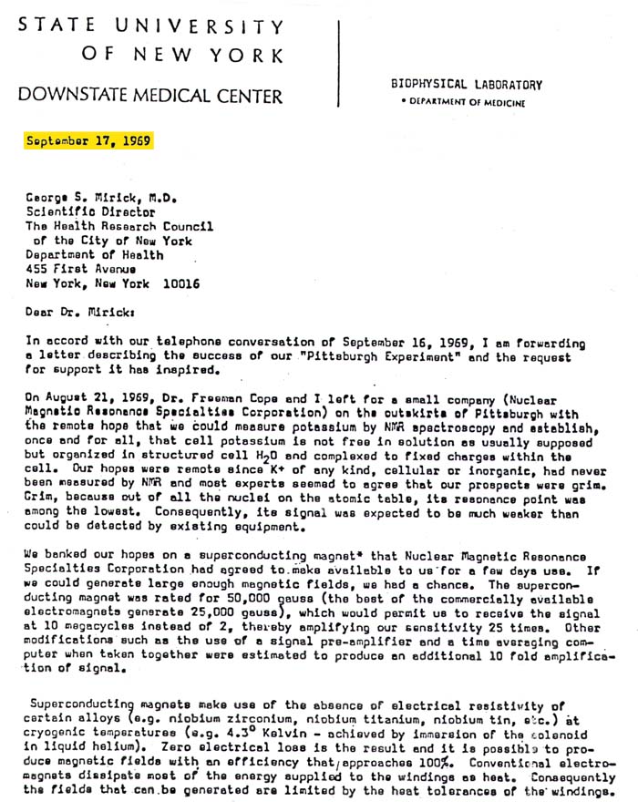
Prior to Damadian's genesis of the NMR body scanning IDEA in 1969, (Figs. 1, 2, 3) NMR instruments for obtaining the NMR spectra of test tube samples had been in operation for 23 years. Thousands of research scientists and chemists the world over used NMR spectrometers to obtain spectra of chemical samples, (Fig 18) but the IDEA to use NMR to scan the human body occurred to no one.
For example, without the IDEA to create an independent self-contained transportable COMBUSTION ENGINE, there would be no AUTOMOBILE. Without the IDEA to transport vehicles by air flotation, there would be no AIRPLANES.
Without the IDEA to generate light by electricity, there would be no LIGHT BULBS and without the IDEA to scan the human body by NMR (Figs. 1, 2, 3) there WOULD BE NO MRI. The IDEA to scan the HUMAN BODY BY NMR ORIGINATED WITH DAMADIAN.
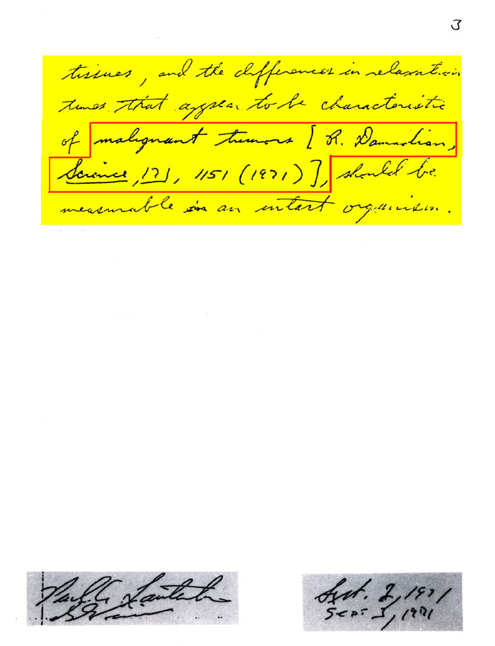
Indeed, when Damadian first originated the IDEA it was ridiculed at a major scientific conference held at the National Cancer Institute of the National Institutes of Health in 1976. A distinguished member of the NMR profession and a professor at the Johns Hopkins University at the time presented his successful repeat of Dr. Damadian's NMR measurements of malignant tissue. He went on to report that other non-malignant diseased tissues also had elevated NMR relaxations. Overlooking that both malignant tissues and non-malignant diseased tissues had marked NMR relaxation elevations when compared to normal that would generate marked differences in pixel brightness (contrast) when displayed on an MRI image he announced.
the human body
by NMR is visionary nonsense." (Fig. 19b)
At another NMR conference that Dr. Damadian was asked to present at, the Moderator of the NMR conference stood up after his presentation and asked
spin the patient ?" (Fig. 19c)
The focused rf of U.S. Patent 3,789,832 (Fig. 20c, 20e) together with shaping of the static magnetic field provided the means for the spatial localization needed for the in vivo scanning of the 106 (Fig. 21 & Fig.22) anatomic loci (pixels – see Fig. 10) that provided the first MR image of the human body. It was demonstrated at trial that the magnetic component (the "near field" that generates the NMR signal) of the transmitted rf (radio frequency) could be shaped to any dimension desired ("focused") and moved to any location desired1. The focusing of the transmitted rf would be used to systematically generate the NMR signals from spatially localized scanning loci within the body, in order to map the anatomy and detect diseased loci.
 Except
for Damadian's BOLD
idea to take
the NMR test-tube analyzer and its 2 1/4 inch sample
space
Except
for Damadian's BOLD
idea to take
the NMR test-tube analyzer and its 2 1/4 inch sample
space
(Fig 18) and totally remake the magnet and its millimeter
diameter NMR rf receiver coils*
that had been devoted unchanged for more than 23
years, (since the inception of NMR), to the chemical
analysis of test-tube samples, there would be no
MRI today.
Notwithstanding grave doubts that there would be sufficient NMR signal/noise reception to detect a tiny NMR signal from a small voxel (volume element) deep within the interior of the human body, using a receiver coil 10 inches away from the signal generating volume, and still achieve an NMR signal large enough to scan the entire human body, the initiative for the NMR body scanner (MRI) proceeded under the Good Lord's protection and direction (Col. 2:3).
It took Damadian to envision the radically transforming step of taking the traditional 23 year old NMR test-tube analyzer and using it to invent a bold new revolutionary scanner for the entire human body.
*rf transmitter coils, rf receiver coils, pre-amplifiers, rf tuned resonance circuits, etc that had been devoted unchanged for more than 23 years since the inception of NMR to the NMR chemical analysis of test-tube samples.
1: see Fig.32 legend.
DISCOVERY OF THE NMR SIGNAL DIFFERENCES TO MAKE THE IMAGE
 Having
originated the NMR body scanner IDEA,
Damadian also provided the means to bring it about.
He isolated the T1 and T2 tissue NMR relaxation
differences (See "T1 and T2 Imaging" section
above. Click here) that made
it possible. He demonstrated for the first time
that the NMR signal
from cancer tissue was markedly different from the
NMR signals obtained
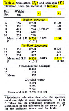
from normal tissues. He further demonstrated in
the same 1971 publication in Science (Damadian,
R. Tumor Detection by Nuclear Magnetic Resonance.
Science, 171:1151-1153, 1971) that the NMR signals
of the normal tissues also differed markedly.
Having
originated the NMR body scanner IDEA,
Damadian also provided the means to bring it about.
He isolated the T1 and T2 tissue NMR relaxation
differences (See "T1 and T2 Imaging" section
above. Click here) that made
it possible. He demonstrated for the first time
that the NMR signal
from cancer tissue was markedly different from the
NMR signals obtained
from normal tissues. He further demonstrated in
the same 1971 publication in Science (Damadian,
R. Tumor Detection by Nuclear Magnetic Resonance.
Science, 171:1151-1153, 1971) that the NMR signals
of the normal tissues also differed markedly.
|
Figure 14 is an axial (cross-sectional) image of the brain showing a tumor of the cerebellum (white areas) in the midline. Figure 14c is a magnified image showing the picture elements or "pixels" (small squares) that make up the image. The cerebellar tumor as it would appear (D) with no MR signal differences. Figure D is the same image as B but where all MR signal differences were eliminated and all the MR pixels therefore had the same pixel brightness. The absence of the MR signal differences between cancer and normal tissue discovered by Damadian gives the MR image pixels equal brightness and the tumor becomes Invisible. |
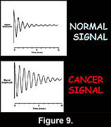
Were the amplitudes of the NMR signals (fig.9) used to set the brightness of each MRI image pixel the same for all tissues (and prior to Dr. Damadian's discovery such NMR tissue signal differences were not known to exist) the brightness of each image pixel would be the same. The MR image would be a blank.
The NMR signal differences discovered by Damadian (Figs 6,9,tables 1 & 2) vary the brightness of the pixels that make up the image (Figs. 6,9). The signal differences of diseased and normal tissues generate the large differences in pixel brightness that enable all diseased tissues (cancerous as well as non-cancerous) to be exquisitely visualized (fig.3b,10,11-13) by the MRI image. Additionally the exceptional NMR signal differences among the normal tissues discovered by Damadian give rise to the extraordinary detail of normal anatomy visualized by MRI (figs. 7,8)
Except for the
marked NMR signal
differences between diseased tissues and normal
tissues discovered
by Damadian and the marked differences of the NMR
signals within
the normal tissues themselves discovered
by Damadian there would be no MRI. It is the tissue
NMR signal differences
discovered by
Damadian that make the MRI image.
The tissue MR (NMR) signal
differences discovered
by Damadian are the BUILDING BLOCKS from which the
MRI IMAGE is CONSTRUCTED.
Without the BUILDING BLOCKS there is no BUILDING!
Without the tissue NMR signal
differences needed to construct an image, that were
discovered by Damadian
(and EXCLUDED by the NP),
THERE IS NOTHING TO MAKE THE IMAGE WITH
and
THERE IS NO MRI!
Except
for the NMR signal
differences discovered
by Damadian to
MAKE THE IMAGE
THERE IS NO IMAGE !
If the NMR signals used to set the brightness of the image pixels do not vary from diseased to normal tissues or within the normal tissues themselves, as discovered by Damadian*, then THERE IS NOTHING TO MAKE THE MRI IMAGE WITH! The
image pixels that the MR scanner |
The cerebellar tumor as it would appear (D) with no MR signal differences. Figure D is the same image as B but where all MR signal differences were eliminated and all the MR pixels therefore had the same pixel brightness. The absence of the MR signal differences between cancer and normal tissue discovered by Damadian gives the MR image pixels equal brightness and the tumor becomes Invisible. |
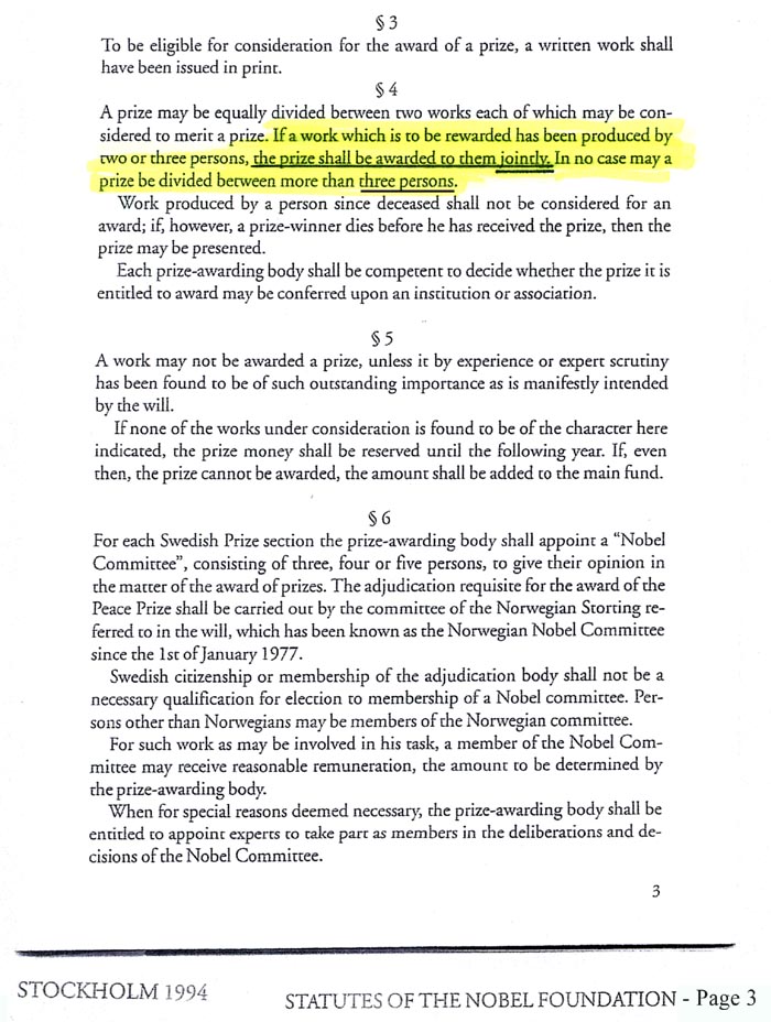
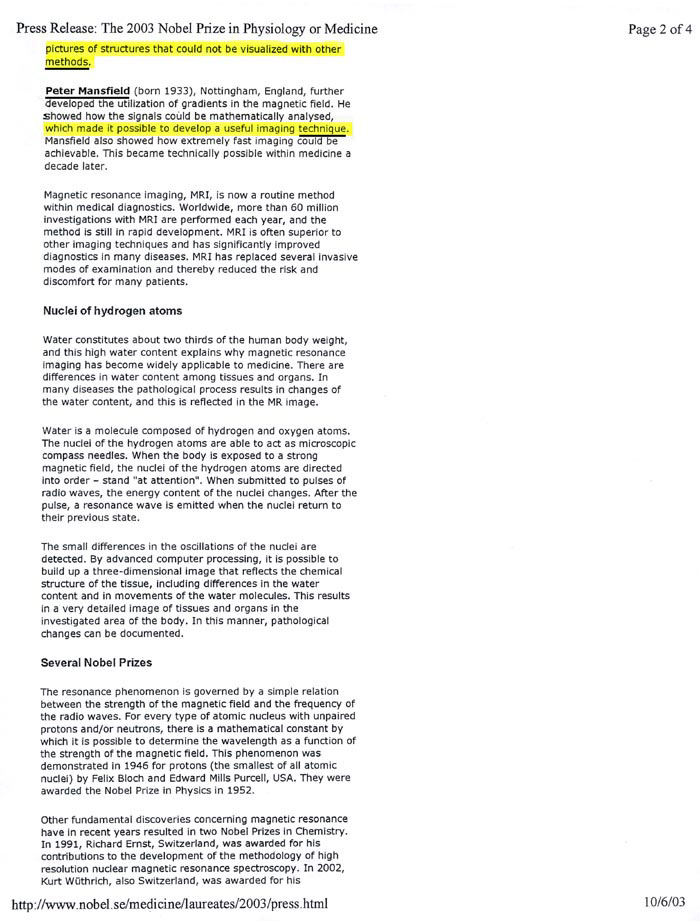
"If a work which is to be rewarded has been produced by two or three persons, the prize shall be awarded to them JOINTLY. In no case may the prize be divided between more than THREE PERSONS." (Nobel Statutes)
The 2003 Nobel Prize for MRI, however was awarded to two persons. P.C. Lauterbur and P. Mansfield, not to the three persons provided for by the Nobel Statutes. The Third Nobel award provided for, and used many times throughout the history of the Nobel Prize, was intentionally VOIDED.
Dr. Damadian was thereby EXCLUDED by the NOBEL (and therefore from history) from any recognition for his critical role in the genesis of the MRI without which MRI WOULD NOT EXIST !
Thus Dr. Damadian's absence from the Nobel Prize for MRI was not an oversight. He was
EXCLUDED INTENTIONALLY
Remarkably, EXCLUDING Dr. Damadian constitutes EXCLUDING THE ONE ORIGINATOR WITHOUT WHOM THERE IS NO MRI AT ALL !! It constitutes EXCLUDING the person who for the first time ever in history conceived of the idea to use a 23 year old test-tube analyzer (Fig. 18, the NMR test-tube analyzer) to scan the live human body, an idea branded as "visionary nonsense" at the time it was originated (Fig. 19b). It also constitutes EXCLUDING the person who further, for the first time ever, PROVIDED the MEANS to ACHIEVE AN MRI IMAGE.
HE PROVIDED THE SIGNAL TO MAKE THE IMAGE WITH (Figs. 4a, Tables I & II, Fig. 4b, Fig. 6) and WITHOUT WHICH THERE IS NO MRI IMAGE AT ALL !!
It is indisputable that Dr. Damadian is the vital contributor by whom (in accordance with the Nobel Statutes) "the work which is to be awarded has been produced."
Indeed as Professor Henry Wallman, Professor of Mathematics, Massachusetts Institiute of Technology (MIT) stated, "I am of the definite opinion that Dr. Damadian's contribution was both prior to and more fundamental than Dr. Lauterbur's" (underlining included in original [ J. Mattson and M. Simon "The Pioneers of NMR and Magnetic Resonance in Medicine: The Story of MRI" Bar-Ilan University Press (1996), 676 ].
Notwithstanding, Dr. Damadian's vital role in the genesis of MRI and the specific provision of the Nobel Statues to provide the award to up to "THREE PERSONS" "JOINTLY", the Nobel Committee intentionally subverted this option (of the Nobel Statutes) and EXCLUDED Dr. Damadian. Remarkably in VOIDING the third award provided by the Nobel Statutes the Nobel Committee intentionally and untruthfully EXCLUDED the only MRI innovator without whom THERE IS NO MRI AT ALL! They EXCLUDED the innovator who originated the idea (Fig. 17d) of the MR body scanner for medical disease and who proposed FOR THE FIRST TIME EVER that the 23 year old non-invasive NMR analyzer of test-tube samples could be used to scan the live human body to detect disease and without which idea no such scanner could ever come into existence. They also EXCLUDED the only one who DISCOVERED the NMR SIGNAL that could achieve it, the NMR SIGNAL that makes the image and without which THERE IS NO IMAGE!
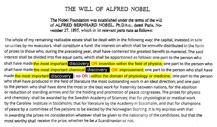
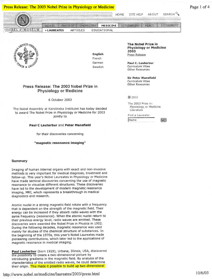
ALFRED NOBEL
For his prize in physics, Nobel specifies it is to be given for the "most important "discovery OR invention". For his prize in chemistry, Nobel specifies it is to be given for the "most important chemical discovery OR improvement".
In so doing, Nobel defines what a discovery is according to HIS WILL.
It is NOT a method !
DELIBERATE VIOLATION
OF
ALFRED NOBEL'S WILL !
the 2003
Nobel Committee
REMARKABLY
EXCLUDED
the only
GENUINE
DISCOVERY
that
QUALIFIED
for
the NOBEL PRIZE
in MEDICINE
under Alfred Nobel's WILL,
Dr. Damadian's DISCOVERY
of the
abnormal NMR signals of
diseased tissue
THAT
MAKE THE IMAGE,
while granting the NOBEL AWARD
INSTEAD to two (METHODS)
that
DID
NOT QUALIFY
for the Nobel Prize in
MEDICINE under Alfred Nobel's WILL1
1 The innovations of METHODS that QUALIFY for the NOBEL PRIZE in Physics and in Chemistry under Alfred Nobel's WILL were explicitly EXCLUDED by Nobel in his WILL for his AWARD in MEDICINE which states that his award in medicine is to be given for
DISCOVERY ONLY.
As Nobel specifies in his WILL his AWARD in MEDICINE shall be given
"to the person who shall have made the most important
DISCOVERY (no OR)
within the domain of physiology or MEDICINE " (ONLY)
in contrast to Nobel's award in physics which states
"to the person who shall have made the most important
DISCOVERY OR Invention
within the field of physics"
and in contrast to Nobel's award in chemistry which states
"to the person who shall have made the most important chemical
DISCOVERY OR Improvement ".
IN
ORDER TO CIRCUMVENT
NOBELS's
EXCLUSION of METHODS
from the NOBEL
PRIZE in MEDICINE
the 2003 NOBEL Committee
FALSELY
DESIGNATED
the Lauterbur and Mansfield
contributions as "discoveries"
(i.e.
their Nobel citation -
"for their discoveries
concerning magnetic resonance imaging")
when they were
not "discoveries"
at all, but
were EXCLUSIVELY
METHODS1,2
(and ONLY
METHODS)
that
DID NOT QUALIFY
for the Nobel Prize in Medicine under Alfred
Nobel's WILL.
They were not
GENUINE DISCOVERIES
of a new scientific phenomenon
such as
Dr. Damadian's
GENUINE
NEW SCIENTIFIC DISCOVERY
,for
the first time ever,
of the
PREVIOUSLY UNKNOWN
ABNORMAL NMR SIGNALS
of
DISEASED TISSUE
(and
their large variations among healthy tissues)
that are used to
GENERATE ALL OF TODAY'S
MRI IMAGES
(and
WITHOUT WHICHTHERE
WOULD BE NO MRI IMAGE AT ALL
!)
They
were, instead,
EXCLUSIVELY
METHODS
2
created to generate
pictures of the abnormal NMR signals of diseased
tissue
GENUINELY
DISCOVERED
by
Dr. Damadian
which SIGNALS
ARE USED
TO MAKE
all of
TODAY'S
MRI IMAGES !
THUS,
in SUMMATION
the
2003 Nobel Committee
SPECIFICALLY
EXCLUDED the
only innovation
that
QUALIFIED
for
the Nobel Prize
in Medicine under Alfred
Nobel's WILL while they made the award
instead to
method innovations 2
EXPLICITLY
EXCLUDED
by Alfred Nobel from the
Nobel Prize in Medicine !
2
Lauterbur provided the METHOD
of using a series of magnetic field gradients
to generate a picture of the abnormal NMR signals
of diseased tissues DISCOVERED
by Damadian.
Mansfield provided ONE
of the METHODS
of making a picture from the spin echo of the
NMR signal, the Echo Planar Image, of the abnormal
NMR signals of diseased tissues DISCOVERED
by Damadian.
 Apart from Dr. Damadian's
ORIGINATION
of the
IDEA*
to take a 23 year old 2¼ inch test-tube analyzer (Fig 18) and REDESIGN AND TRANSFORM IT into a non-invasive scanner for detecting disease in the live human body (Figs 1,2,3,20e)
and
Apart from Dr. Damadian's
DISCOVERY**
of the
disease detecting signal
to do it with
(Figs 3,4a,4b,9,11: tables 1 & 2)
THERE WOULD BE NO MRI TODAY !!
(Fig 22, Fig 11)
(The Economist, Dec. 4, 2003 (Q4 2003))
* An IDEA publically branded "NONSENSE" at the time (Cursor click on the black rectangle of interest to visit the cited reference)
** ... the discovery that tissue disease altered the NMR radio signal produced by the body's tissues (Figure 9). The diseased tissue NMR radio signals obtained in an NMR scan of the live human body were thereby made distinct (Figure 9) from the tissue NMR radio signals obtained from the bodies surrounding normal tissues (Figure 11) . The discovery enabled detailed visualization of the bodies' diseased tissues (Figure 11) for the first time in medical history ever thereby making possible NMR (MRI) scans of the live human body (enabling early treatment).
Without Edison there is no Light Bulb
Without Alexander Graham Bell there is no Telephone
Without the Wright Brothers there is no Airplane
Without Damadian there is no MRI*
*as affirmed by
TWO PRESIDENTS OF THE UNITED STATES,
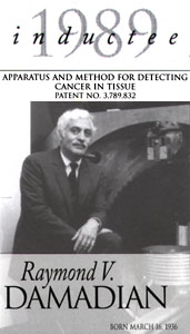
THE NATIONAL INVENTORS HALL OF FAME,
THE UNITED STATES SUPREME COURT.
THE TRUTH WILL OUT"
William Shakespeare (1598 AD)
"The Merchant of Venice"
Launcelot Gobbo to Old Gobbo (Launcelot's father)
the
Nobel Committee's effort
to
erase Dr. Damadian
from the history of his invention and from his
7 years
of suffering to surmount the
"INDOMITABLE"
technological obstacles
to convert
a 10 mm test-tube analyser
into a scanner of the LIVE human body
what the Nobel Committee appears not to appreciate, in contrast to
Launcelot Gobbo,
is that
IS
INVINCIBLE !!
FONAR Position Regarding Nobel Prize
Dr.
Damadian Interview - online
(requires a computer with a sound card)
The Economist celebrates innovation at the 2nd Annual Innovation Awards and Summit in San Francisco

<
more on Fonar's Stand-Up MRI >
![]()
Site
Map | Terms
of Use-Our Privacy Policy Use
Copyright © 2003-20014 FONAR- All Rights Reserved